Please note
This trial is no longer recruiting patients. We hope to add results when they are available.
Brain (and spinal cord) tumours
Closed
Other
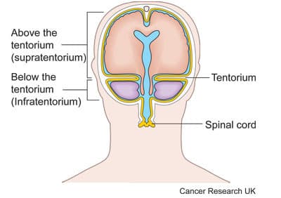
This study is to see if a PET-CT scan and magnetic resonance spectroscopy (MRS) can help doctors make an assessment of brain tumours. The study is open to people with a type of brain tumour called a glioma that is above the hind brain (cerebellum) and brain stem. Doctors call this a supratentorial glioma.
Doctors use magnetic resonance imaging (MRI) scans to diagnose brain tumours. They are best at finding out the size of the tumour and where it is. But they don’t give doctors all the information they would like.
Magnetic resonance spectroscopy (MRS) scans are similar to MRI scans, but can give extra information. MRS scans may be able to give information about how quickly your tumour is growing and if certain treatments will work.
In this study, they are also looking at PET-CT scans. This scan combines a PET scan and CT scan into one scan.
Before having a PET-CT scan, you have an injection of a small amount of radioactive drug called a tracer. In this study the researchers are using a tracer that may give doctors more information about your tumour and how fast it is growing.
The researchers will compare the results of these 2 scans with other tests done on a sample of your tumour tissue. The main aim is to find out how good MRS and PET-CT scans are for assessing gliomas.
Recruitment start: 18 December 2014
Recruitment end: 31 March 2016
Please note: In order to join a trial you will need to discuss it with your doctor, unless otherwise specified.
Dr Adam Waldman
Imperial College London
NIHR Biomedical Research Centres (BRCs) Award
NIHR Clinical Research Network: Cancer
Last reviewed: 30 Mar 2016
CRUK internal database number: 12771
About Cancer generously supported by Dangoor Education since 2010. Learn more about Dangoor Education


Search our clinical trials database for all cancer trials and studies recruiting in the UK.
Connect with other people affected by cancer and share your experiences.
Questions about cancer? Call freephone 0808 800 40 40 from 9 to 5 - Monday to Friday. Alternatively, you can email us.