Radiotherapy
When you have surgery your surgeon sends what they remove for various tests in a laboratory. These tests help them work out the risk of your cancer coming back.
For intermediate risk womb cancer, you may have internal radiotherapy when your cancer is:
stage 1A grade 3
stage 1B grade 1 to 2
stage 2 grade 1
For high-intermediate womb cancer, your doctor may sometimes consider internal radiotherapy instead of external radiotherapy. This is usually when the risk is lower, and lymph nodes that were removed during surgery are clear of cancer.
You may have up to 3 treatments lasting about 20 minutes each.
If you can’t have surgery or if you’re not fit enough to have surgery, you may have internal radiotherapy:
on its own for a low grade cancer
after external radiotherapy for a high grade cancer
Depending on your situation, you may have 3 to 6 treatments lasting about 20 minutes each.
You have treatment in the radiotherapy department. Depending on the type of brachytherapy you have, you might stay in hospital to have it or attend as an outpatient. You usually have treatment as an:
inpatient if you haven't had a hysterectomy
outpatient if you've had a hysterectomy
You go into hospital before your brachytherapy. The nurses take you to the theatre to have an anaesthetic. The anaesthetic may be an injection into your spine (epidural) so you are numb below the waist. Or you have a general anaesthetic, which puts you to sleep.
The doctor puts the applicators into the vagina and cervix. The applicators are made up of tubes and, or needles. The doctor places gauze inside the vagina to hold the applicators in place. They also put a tube (catheter) into your bladder.
Once the applicators are in place, you cannot leave the bed and must remain flat. Below is a diagram of how the applicators are placed.

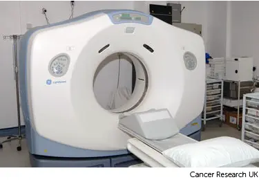
You have a CT and, or MRI scan to check the position of the applicators. Below is a photograph of a CT scanner.
The nurses on the ward make sure you are comfortable. You can have pain relief if you need it. You remain on the ward while the treatment team produce a treatment plan. This should be ready within a few hours.
Once the plan is ready you come down from the ward to the brachytherapy room. The radiographers connect the applicators to the machine. During the treatment, they leave the room and watch you from outside on a CCTV screen.
Afterwards, you might have light sedation for the removal of the applicators and bladder catheter. You can then go back to the ward and can usually go home on the same day.
It is important that another adult takes you home. They should also stay with you for 24 hours afterwards. For 24 hours after a general anaesthetic, you should not:
drive a car
operate machinery
drink alcohol
You should not feel unwell after treatment, but you may notice some:
slight bleeding from your vagina
mild period like cramps
This treatment involves placing a tube inside the vagina. Your doctor takes it out once the treatment is over. You are only radioactive when the treatment machine is switched on. So afterwards you are safe to be around everyone, including children.
A special brachytherapy machine in a purpose built room gives the treatment. You won’t feel the radiotherapy, but the tube can feel uncomfortable.
When you arrive for treatment, the radiographers may ask you to empty your bladder. Your doctor or therapy radiographer examines you to check what size applicator they can use for the treatment. The applicator is a tube which comes in different sizes. They put the tube in your vagina and hold it in place with a clamp. They use a gel to help put the applicator in so it’s as comfortable as possible. It should not be painful.
You then have a CT scan, which takes a short time. Your radiographers wait outside while this happens. Below is a photograph of a CT scanner.
Your radiographers remove the applicator after the scan and you are free to go home.
Your radiographers and doctors create your radiotherapy plan. They ensure that the area of the cancer will receive a high dose and surrounding areas receive a low dose. This reduces the side effects you might get during and after treatment.
You often have your first treatment on the same day but may need a more individualised plan. In this case, you will come back on another day for your first treatment.
You usually come back for treatment within a week. You stay in the same position as you were for the CT scan. Your radiographers put in the applicator and connect it to the brachytherapy machine. They then leave the room but can still see you on a CCTV screen during treatment.
Your radiographers remove the applicator once treatment has finished. You are then free to go home. The process is the same every time.
You should not feel unwell after treatment, but you may notice some:
slight bleeding from your vagina
mild period like cramp
You might have to travel a long way each day for your radiotherapy. This depends on where your nearest cancer centre is. This can make you very tired, especially if you have side effects from the treatment.
You can ask your radiographers for an appointment time to suit you. They will do their best, but some departments might be very busy. Some radiotherapy departments are open from 7 am till 9 pm.
Car parking can be difficult at hospitals. Ask the radiotherapy staff if you are able to get free parking or discounted parking. They may be able to give you tips on free places to park nearby.
Hospital transport may be available if you have no other way to get to the hospital. But it might not always be at convenient times. It is usually for people who struggle to use public transport. Or who have any other illnesses or disabilities. You might need to arrange hospital transport yourself.
Some people are able to claim back a refund for healthcare travel costs. This is based on the type of appointment and whether you claim certain benefits. Ask the radiotherapy staff for more information about this and hospital transport.
Some hospitals have their own drivers and local charities might offer hospital transport. So do ask if any help is available in your area.
Radiotherapy can cause different side effects including:
diarrhoea
bladder problems such as a burning feeling when you pass urine or passing urine more often
a sore vagina, and vaginal discharge or bleeding
In the long term, your vagina may become shorter, narrower and less stretchy. To try to prevent this, your radiographer or nurse will give you vaginal dilators to use regularly after your radiotherapy treatment.
Last reviewed: 12 Apr 2024
Next review due: 12 Apr 2027
Side effects of internal radiotherapy tend to happen about 1 to 2 weeks after treatment. They are usually mild and last for a few days or weeks.
Radiotherapy uses high energy x-rays to treat womb cancer cells. You have the treatment in the hospital radiotherapy department.
Most women with womb cancer have surgery to remove the womb. The operation you have depends on how far the cancer has grown.
After treatment for womb cancer, you have checkups at the hospital. You also have tests, including blood tests, x-rays and scans.
There is support available during and after treatment to help you cope. This includes support from your clinical nurse specialist, cancer charities, community services, and family and friends.
The womb is the pear shaped muscular bag that holds a baby during pregnancy. Most womb cancers start in the lining of the womb. They are also called uterine or endometrial cancer.
About Cancer generously supported by Dangoor Education since 2010. Learn more about Dangoor Education


Search our clinical trials database for all cancer trials and studies recruiting in the UK.
Connect with other people affected by cancer and share your experiences.
Questions about cancer? Call freephone 0808 800 40 40 from 9 to 5 - Monday to Friday. Alternatively, you can email us.