Tests and scans
MRCP stands for magnetic resonance cholangio pancreatography (col-an-jee-oh pan-kree-at-og-raf-ee). An MRCP scan is a type of MRI scan that you have in an MRI scanner.
MRI stands for magnetic resonance imaging. An MRI scan produces pictures from angles all around the body and shows up soft tissues very clearly.
These scans create pictures using magnetism and radio waves to give detailed pictures of your:
pancreas
gallbladder
bile ducts
liver
You might have an MRI scan of your tummy (abdomen) at the same time as the MRCP.
A operates the scanner and looks after you while you have your scan. You usually have these scans in the x-ray (radiology) department as an outpatient. It can take up to 60 minutes. Sometimes it takes longer depending on the preparation you need.
MRCP isn't available in all hospitals.
You might have one or both of these scans to find out:
if you have any abnormal areas in your abdomen
the size of the cancer if you have been diagnosed with cancer, and whether it has spread
Before you go to your appointment, or when you arrive, you fill in a safety checklist. This asks about:
any operations you’ve had
whether you have any metal implants or other metals in your body
An MRI scan uses strong magnetism which could affect any metal in your body. These include:
pacemakers or an implantable defibrillator (to treat abnormal heart rhythms)
surgical clips, pins or plates
cochlear implants (for deafness)
metal fragments anywhere in your body – for example from an injury, dental fillings and bridges
You can have an MRI scan if you have some metals in your body. But your doctor and radiographer decide if it is safe for you. Tell your radiographer about any metals in your body.
Some stick-on medicine patches contain metal and could overheat in the MRI scan, causing burns. Tell your radiographer before the scan if you use medicine patches. You might need to remove them before the scan.
It is important to let your radiographer know if you have any tattoos. This is because some tattoo ink contains metal.
Some people feel claustrophobic or closed in when they’re having an MRI scan. Contact the department before your test if you’re likely to feel like this. The hospital staff can take extra care to make sure you’re comfortable and that you understand what’s going on. Your doctor can give you medicine to help you relax if you need it.
Tell the department staff beforehand if you are pregnant or think you might be pregnant. An MRI is generally safe during pregnancy. But as a precaution, you usually won’t have one during the first 3 months of your pregnancy.
You might need to stop eating and drinking for a few hours before an MRCP scan. If you are having an abdominal (tummy) MRI scan at the same time, you might need to stop eating and drinking around 4 to 5 hours beforehand. This varies between hospitals, so do check your appointment letter for more information.
Talk to your doctor if not eating and drinking before the test could be a problem, for example, if you are diabetic.
Let the staff in the MRI department know what medicines you are taking. They will tell you if you should stop taking any of them before your scan.
When you arrive at the scanning department, the radiographer might ask you to change into a hospital gown. You might not have to undress if your clothing doesn’t have any metal, such as zips or clips.
You have to:
remove any jewellery, including body piercings and your watch
remove hair clips
empty your pockets of coins and keys
It’s safe to take a relative or friend into the scanning room with you. But check with the department staff first. Your friend or relative will also need to remove any metal they have on them.
Once ready your radiographer takes you into the scanning room.
The MRI machine is large and shaped like a doughnut.
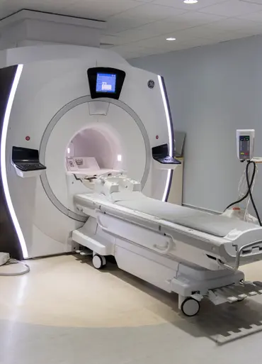
You lie on your back on a couch that can slide into the MRI machine. The radiographer places a light piece of equipment known as the coil to rest on top of your abdomen. This helps to collect information as you have the scan.
You might have an injection of a dye (contrast medium) through a into a vein in your arm. The dye helps to show up your body’s organs more clearly. Some people are allergic to the dye. Your radiographer will check first about any medical conditions or allergies you have.
After the dye injection you may:
feel sick
have a headache
feel warm or flushed
have a metallic taste in your mouth
feel a little dizzy
These effects are usually mild and last for a short time. Tell your radiographer if you feel unwell at any point during or after your scan.
You need to lie as still as possible. The scan is painless but it can be uncomfortable to stay still. Tell the radiographer if you're getting stiff and need to move.
Once you’re in the right position on the couch, your radiographer leaves the room. They can see you on a TV screen or through a window at all times from the control room. You can talk to each other during the scan, usually through an intercom. You will have a button you can press if you want them to stop the scan immediately if you feel claustrophobic.
The couch moves through the MRI scanner. It takes pictures as you move through it. Your radiographer might ask you to hold your breath at times.
The scanner makes a very loud clanging sound throughout the scan. You wear headphones or earplugs to protect your hearing. You can also listen to music. Keeping your eyes closed can help.
This 1 minute video shows you what happens when you have an MRI scan.
When the scan is over, your radiographer comes back into the room and lowers the couch so that you can get up.
You usually stay in the department for about 15 to 30 minutes after your scan if you've had the dye. In that time, the scanning staff check that you are not feeling unwell.
Your radiographer removes the cannula from the vein in your arm before you leave.
You should then be able to go home or back to work, and also eat and drink normally.
An MRCP and MRI are very safe and don’t use radiation. Some people can’t have an MRCP or MRI, but the checklist picks this up beforehand. Your doctor and radiographer make sure the benefits of having the test outweigh any possible risks.
Some of the possible risks include:
You might get a small bruise around the area where they put the needle in for the cannula.
There's a risk that the contrast medium will leak outside the vein. This can cause swelling and pain in your arm but it’s rare. Tell your radiographer if you have any swelling or pain. Let your GP know if it doesn’t get better or starts to get worse when you’re at home.
An allergic reaction to the contrast medium injection is rare. This most often starts with feeling weak, sweating and difficulty breathing. Tell your radiographer straight away if you feel unwell so they can give you medicine to control the reaction.
You should get your results within 1 or 2 weeks.
Ask your doctor, radiographer or nurse how long it will take to get them. Contact the doctor who arranged the test if you haven’t heard anything after a couple of weeks.
Waiting for test results can be a very worrying time. You might have contact details for a specialist nurse. You can get in touch with them for information and support if you need to. It may help to talk to a close friend or relative about how you feel.
We have more information on tests, treatment and support if you have been diagnosed with cancer.
Last reviewed: 17 Jul 2025
Next review due: 17 Jul 2028
An MRI scan creates pictures using magnetism and radio waves. Find out why you might have it, how you have it and the possible risks.
Search for the cancer type you want to find out about. Each section has detailed information about symptoms, diagnosis, treatment, research and coping with cancer.
Find out about tests to diagnose cancer and monitor it during and after treatment, including what each test can show, how you have it and how to prepare.
Gallbladder cancer is a cancer that begins in the gallbladder, which is part of the biliary system. It is quite rare in the UK and more common in women than in men.
About Cancer generously supported by Dangoor Education since 2010. Learn more about Dangoor Education


Search our clinical trials database for all cancer trials and studies recruiting in the UK.
Connect with other people affected by cancer and share your experiences.
Questions about cancer? Call freephone 0808 800 40 40 from 9 to 5 - Monday to Friday. Alternatively, you can email us.