Treatment for neuroendocrine cancer
Read more about the difference between NETs and NECs
Your treatment plan depends on what type of neuroendocrine cancer you have.
You might have external radiotherapy to control symptoms if your NET has spread. For example, you might have radiotherapy to control pain caused by cancer that spreads to your bones.
You might have external radiotherapy for some types of NEC. You might have radiotherapy:
combined with - this is called chemoradiotherapy
after surgery - this is called adjuvant radiotherapy
to control symptoms of advanced cancer - this is called palliative radiotherapy
The radiotherapy team plan your external radiotherapy before you start treatment. This means working out the dose of radiotherapy you need and exactly where you need it.
Your planning appointment takes from 15 minutes to 2 hours.
You usually have a planning in the radiotherapy department.
The scan shows the cancer and the area around it. You might have other types of scans or x-rays to help your treatment team plan your radiotherapy. The plan they create is just for you.
Read more about radiotherapy planning
Radiotherapy machines are very big and could make you feel nervous when you see them for the first time. The machine might be fixed in one position. Or it might rotate around your body to give treatment from different directions. The machine doesn't touch you at any point.
Before your first treatment, your will explain what you will see and hear. In some departments, the treatment rooms have docks for you to plug in music players. So you can listen to your own music while you have treatment.
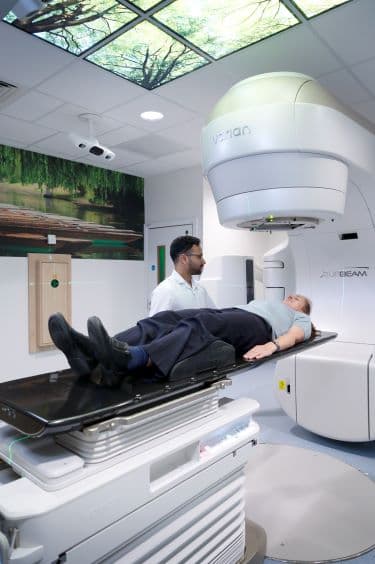
You need to lie very still. Your radiographers might take images (x-rays or scans) before your treatment. This is to make sure that you're in the right position. The machine makes whirring and beeping sounds. You won’t feel anything when you have the treatment.
Your radiographers can see and hear you on a CCTV screen in the next room. They can talk to you over an intercom and might ask you to hold your breath or take shallow breaths at times. You can also talk to them through the intercom or raise your hand if you need to stop or if you're uncomfortable.
This type of radiotherapy won't make you radioactive. It's safe to be around other people, including pregnant women and children.
You might have to travel a long way each day for your radiotherapy. This depends on where your nearest cancer centre is. This can make you very tired, especially if you have side effects from the treatment.
You can ask your radiographers for an appointment time to suit you. They will do their best, but some departments might be very busy. Some radiotherapy departments are open from 7 am till 9 pm.
Car parking can be difficult at hospitals. Ask the radiotherapy staff if you are able to get free parking or discounted parking. They may be able to give you tips on free places to park nearby.
Hospital transport may be available if you have no other way to get to the hospital. But it might not always be at convenient times. It is usually for people who struggle to use public transport. Or who have any other illnesses or disabilities. You might need to arrange hospital transport yourself.
Some people are able to claim back a refund for healthcare travel costs. This is based on the type of appointment and whether you claim certain benefits. Ask the radiotherapy staff for more information about this and hospital transport.
Some hospitals have their own drivers and local charities might offer hospital transport. So do ask if any help is available in your area.
The side effects depend on which body part you're having treatment to. It also depends on the dose of radiotherapy. Radiotherapy to control symptoms is usually a lower dose. So you are less likely to get side effects.
The most common side effects of radiotherapy during and just after treatment are:
reddening of the skin in the treatment area
tiredness
loss of hair in the treatment area
Read more about radiotherapy side effects
Treatment for neuroendocrine cancer can be difficult to cope with for some people. Your nurse will give you phone numbers to call if you have any problems at home.
Find out more about coping with a neuroendocrine cancer and how to get support
Last reviewed: 23 Mar 2021
Next review due: 22 Mar 2024
Neuroendocrine cancers are also called neuroendocrine neoplasms (NENs). There are 2 key groups - neuroendocrine tumours (NETs) and neuroendocrine carcinomas (NECs).
Treatment depends on the type of neuroendocrine cancer you have, where it is, its size and whether it has spread (the stage).
PRRT is a type of radioisotope therapy for NETs. It is also called radioligand therapy. It uses a radioactive substance. This is attached to a man made form of a hormone called somatostatin.
Selective internal radiation therapy (SIRT) is a type of brachytherapy used to treat cancer in the liver. This is usually for bowel cancer that has spread to the liver (liver secondaries).
Practical and emotional support is available to help you cope with neuroendocrine cancer.
Neuroendocrine cancers develop in cells of the neuroendocrine system. They can develop in different parts of the body including the lungs, stomach, pancreas and bowel.
About Cancer generously supported by Dangoor Education since 2010. Learn more about Dangoor Education


Search our clinical trials database for all cancer trials and studies recruiting in the UK.
Connect with other people affected by cancer and share your experiences.
Questions about cancer? Call freephone 0808 800 40 40 from 9 to 5 - Monday to Friday. Alternatively, you can email us.