Radiotherapy for breast cancer
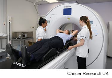
You usually have a planning CT scan in the radiotherapy department.
Your planning appointment takes from 15 minutes to 2 hours.
The scan shows the part of the body that needs treatment. You might have other types of scans or x-rays to help your treatment team plan your radiotherapy. The plan they create is just for you.

Your radiographers tell you what is going to happen. They help you into position on the scan couch. You usually have to undress from the waist up but you can ask for a gown to help maintain your privacy.
You lie on the CT couch on a special board called a breast board. You might also need to raise one or both arms above your head.
It's important to continue the arm exercises you were shown after your surgery. This will make it easier to get your arm in the right position. It will also help prevent your arm and shoulder from becoming stiff during and after treatment.
Find out more about exercises after surgery to the breast
You will lie in the same position on the breast board for both the CT scan and the radiotherapy treatment. You need to lie very still so tell your radiographers if you aren't comfortable. They can help get you into a more comfortable position.
Also let your radiographer know if you think you may be pregnant or have a .
Once you are in position, your radiographers put some markers on your skin. They move the couch up and through the scanner. They then leave the room and the scan starts.
The scan takes about 5 minutes. You won't feel anything. Your radiographers can see and hear you from the CT control room where they operate the scanner.
Your treatment team puts all the scans together in a special computer to decide your radiotherapy plan.
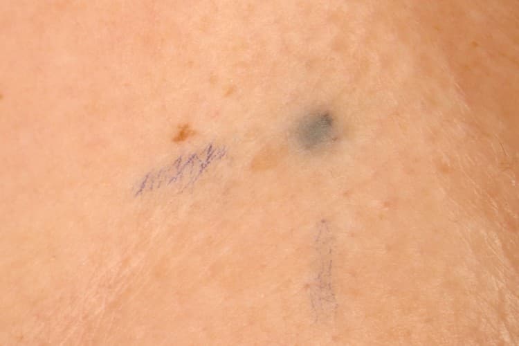
The radiographers may make pin point sized tattoo marks on your skin. They use these marks to line you up into the same position every day. The tattoos make sure they treat exactly the same area for all of your treatments. They may also draw marks around the tattoos with a permanent ink pen so that they are clear to see when the lights are low.

The radiotherapy staff tells you how to look after your skin and the markings. The pen marks might start to rub off in time, but the tattoos won’t. Tell your radiographer if that happens. Don't try to redraw them yourself.
Your treatment team might make a mould for you. They call this a shell.
You wear the shell during the treatment sessions to keep your breasts in the same position each time. The radiographers might also make marks on it. They use the marks to line up the radiotherapy machine for each treatment.
The process of making a shell can vary slightly between hospitals. It usually takes around 30 minutes.
You need to wear clothes that you can easily take off. You also need to take off any jewellery from that area.
A radiographer or technician uses a special kind of plastic that they heat in warm water. This makes it soft and easily bent. They put the plastic onto your chest so that it moulds exactly. It feels a little like a warm flannel.
After a few minutes, the plastic gets hard. The radiographer takes the shell off and it is ready to use.
Breast cancer can sometimes spread to other parts of the body such as the brain. This is secondary or advanced breast cancer. You may have radiotherapy to the brain to help relieve any symptoms you have.
You will have a head mask (or mould) made if you’re having radiotherapy to the brain. This keeps your head still during your planning CT scan and while you have treatment.
Find out more about having radiotherapy to the brain
You might have to wait a few days or up to 3 weeks before you start treatment. During this time radiographers, physicists and your doctor create a precise radiotherapy plan for you.
They make sure the treatment area receives a high dose and nearby areas receive a low dose. This reduces the side effects you might get during and after treatment.
Last reviewed: 29 Jun 2023
Next review due: 29 Jun 2026
You might have external beam radiotherapy after breast surgery to lower the risk of the cancer coming back. This is usually over 1 to 3 weeks depending on your situation.
The side effects of external radiotherapy to the breast include tiredness and changes to the skin in the treatment area.
Deciding about treatment can be difficult when you have secondary breast cancer. Treatments such as chemotherapy or radiotherapy can help to reduce symptoms and might make you feel better.
Get practical, physical and emotional support to help you cope with a diagnosis of breast cancer, and life during and after treatment.
Breast cancer is cancer that starts in the breast tissue. Find out about who gets breast cancer and where it starts.
Find out about breast cancer, including symptoms, diagnosis, treatment, survival, and how to cope with the effects on your life and relationships.

About Cancer generously supported by Dangoor Education since 2010. Learn more about Dangoor Education
Search our clinical trials database for all cancer trials and studies recruiting in the UK.
Connect with other people affected by cancer and share your experiences.
Questions about cancer? Call freephone 0808 800 40 40 from 9 to 5 - Monday to Friday. Alternatively, you can email us.