Radiotherapy
The radiotherapy team plan your external beam radiotherapy before you start treatment. This means working out the dose of radiotherapy you need and exactly where you need it. Your planning appointment can take between 15 minutes and 2 hours.
Read more about why you have radiotherapy
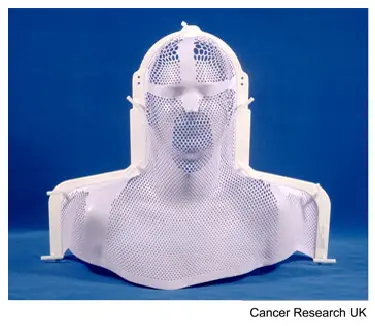
You usually have a mask if you are having radiotherapy for laryngeal cancer. You might also hear this called a mould or shell. The mask keeps you perfectly still while you are having treatment. The radiographers may also make marks on it. They use the marks to line up the radiotherapy machine for each treatment.
The process of making the mask can vary slightly between hospitals. It usually takes around 30 minutes.
The mask is normally made directly against your skin. It's helpful to wear clothing that you can easily take off. You will also need to take off any jewellery from that area.
Having a lot of facial hair can make it difficult to make a head and neck mask. The radiotherapy staff will discuss this with you.
A technician or radiographer makes the mask in the mould room of the radiotherapy department or during your CT planning scan.
The process of making a mask can vary slightly between hospitals. Most often they use a special kind of plastic heated in warm water or an oven that makes it soft and pliable.
Your technician puts the plastic mesh on your face so that it moulds to fit your face exactly. It feels a little like having a warm flannel put onto your face. You can still breathe easily, as the plastic has lots of holes in it. The staff explain what is going to happen.
After a few minutes the plastic mesh becomes hard. Your technician takes the mask off. It is then ready for use.
The video below shows what happens when you have your mesh mask made. The video is about 1 and a half minutes long.
You usually have a planning CT scan in the radiotherapy department.
The scan shows the cancer and the area around it. You might have other types of scans or x-rays to help your treatment team plan your radiotherapy. The plan they create is just for you.
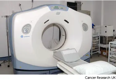
You lie on the scanner couch. Your radiographers will tell you if you need to remove any clothes. They need to see the treatment area, but will cover you up as much as possible. You have the mask in place for your planning scan.
You have to lie as still as you can, so that the measurements are accurate and the radiographers can record your exact position. This means they can make sure you are lying in the correct position every time you have treatment. Tell the radiographers if you are uncomfortable.
Once you are in position, the radiographers move the couch up and through the scanner. The scanner is a doughnut shape. Your radiographers leave the room and the scan starts. It takes about 5 minutes. You won't feel anything. Your radiographers watch from the room next door and you can talk to them on an intercom if you need to.
You might need an injection of contrast dye into a vein in your hand. This is a dye that helps show up body tissues more clearly on the scan.
Before you have the injection of the contrast dye, your radiographer asks you about any medical conditions or allergies. Some people are allergic to the dye.
Watch our video about radiotherapy planning. It is just under 3 minutes long.
When you finish your planning appointment your radiographers will help you off the CT scanner couch. Then you can get changed back into your clothes. You will stay in the department for about 15 to 30 minutes afterwards if you had an injection of the dye. This is in case it makes you feel unwell, which is rare.
You should be able to go home or back to work. You can eat and drink normally.
You might have to wait a few days or up to 3 weeks before you start treatment.
During this time the physicists and your radiotherapy doctor (clinical oncologist) decide the final details of your radiotherapy plan. They make sure that the area of the cancer will receive a high dose and nearby areas receive a low dose. This reduces the side effects you might get during and after treatment.
Last reviewed: 12 Aug 2024
Next review due: 12 Aug 2027
Radiotherapy uses high energy x-rays to treat laryngeal cancer. You have the treatment in the hospital radiotherapy department.
You might have surgery, chemotherapy, radiotherapy or a combination of treatments to treat laryngeal cancer.
Laryngeal cancer is cancer that starts in the voice box (larynx). It is a type of head and neck cancer.
Lots of advice and support are available to help you cope with living with laryngeal cancer.
About Cancer generously supported by Dangoor Education since 2010. Learn more about Dangoor Education


Search our clinical trials database for all cancer trials and studies recruiting in the UK.
Connect with other people affected by cancer and share your experiences.
Questions about cancer? Call freephone 0808 800 40 40 from 9 to 5 - Monday to Friday. Alternatively, you can email us.